


 Foundational characteristics of cancer include proliferation, angiogenesis, migration, evasion of apoptosis, and cellular immortality. Find key markers for these cellular processes and antibodies to detect them.
 The SUMOplot™ Analysis Program predicts and scores sumoylation sites in your protein. SUMOylation is a post-translational modification involved in various cellular processes, such as nuclear-cytosolic transport, transcriptional regulation, apoptosis, protein stability, response to stress, and progression through the cell cycle.
 The Autophagy Receptor Motif Plotter predicts and scores autophagy receptor binding sites in your protein. Identifying proteins connected to this pathway is critical to understanding the role of autophagy in physiological as well as pathological processes such as development, differentiation, neurodegenerative diseases, stress, infection, and cancer.
ERAP2 Antibody
- SPECIFICATION
- CITATIONS
- PROTOCOLS
- BACKGROUND

Application
| WB, E |
|---|---|
| Primary Accession | Q6P179 |
| Other Accession | NP_071745, 11641261 |
| Reactivity | Human |
| Host | Rabbit |
| Clonality | Polyclonal |
| Isotype | IgG |
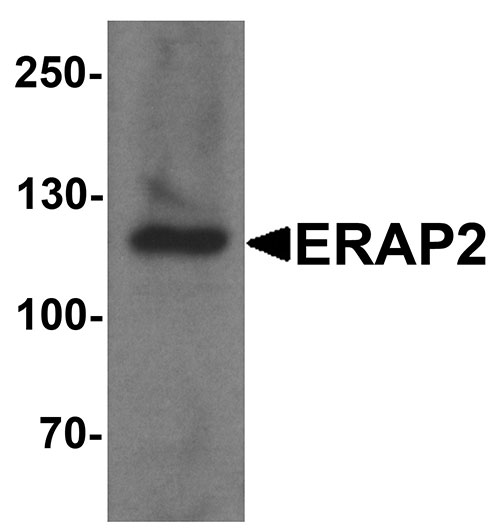
| Calculated MW | Predicted: 106 kDa Observed: 120kDa |
| Application Notes | ERAP2 antibody can be used for detection of ERAP2 by Western blot at 1 - 2 µg/ml. |
| Gene ID | 64167 |
|---|---|
| Target/Specificity | ERAP2; ERAP2 antibody is human reactive. Multiple isoforms of ERAP2 are known to exist. ERAP2 antibody is predicted to not cross-react with ERAP1. |
| Reconstitution & Storage | ERAP2 antibody can be stored at 4℃ for three months and -20℃, stable for up to one year. |
| Precautions | ERAP2 Antibody is for research use only and not for use in diagnostic or therapeutic procedures. |
| Name | ERAP2 |
|---|---|
| Synonyms | LRAP |
| Function | Aminopeptidase that plays a central role in peptide trimming, a step required for the generation of most HLA class I-binding peptides. Peptide trimming is essential to customize longer precursor peptides to fit them to the correct length required for presentation on MHC class I molecules. Preferentially hydrolyzes the basic residues Arg and Lys. |
| Cellular Location | Endoplasmic reticulum membrane; Single-pass type II membrane protein |
| Tissue Location | Ubiquitously expressed. Highly expressed in spleen and leukocytes. |

Thousands of laboratories across the world have published research that depended on the performance of antibodies from Abcepta to advance their research. Check out links to articles that cite our products in major peer-reviewed journals, organized by research category.
info@abcepta.com, and receive a free "I Love Antibodies" mug.

Provided below are standard protocols that you may find useful for product applications.
Background
The endoplasmic reticulum (ER) aminopeptidase 2 (ERAP2), a member of the peptidase M1 family, like the related protein ERAP1, plays a central role in peptide trimming, a step required for the generation of most HLA class I-binding peptides (1,2). Like ERAP1, ERAP2 is localized to the lumen of the ER and is thought to associate with ERAP1 as a heterodimer (1). Both ERAP1 and ERAP2 have been linked to several human diseases ranging from infections to autoimmunity and cancer, and may play a role in the innate immune response (reviewed in 3).
References
Saveanu L, Carroll O, Lindo V, et al. Concerted peptide trimming by EARP1 and ERAP2 aminopeptidase complexes in the endoplasmic reticulum. Nat. Immunol. 2005; 6:689-97.
Saric T, Chang SC, Hattori A, et al. An IFN-gamma induced aminopeptidase in the ER, ERAP1, trims precursors to MHC class I-presented peptides. Nat. Immunol. 2002; 3:1169-76.
Cifaldi L, Romania P, Lorenzi S, et al. Role of endoplasmic reticulum aminopeptidases in health and disease: from infection to cancer. Int. J. Mol. Sci. 2012; 13:8338-52
If you have used an Abcepta product and would like to share how it has performed, please click on the "Submit Review" button and provide the requested information. Our staff will examine and post your review and contact you if needed.
If you have any additional inquiries please email technical services at tech@abcepta.com.
Ordering Information
Other Products
Shipping Information
